Patient information · Assessment and treatment
TMJ condylography in Dubai: recording jaw movement
Condylography records lower-jaw movements as traces and numerical measurements. It complements the clinical examination, during which I assess how freely your jaw moves and which movements are uncomfortable. I use the Gamma CADIAX system in my practice.
What can the recording help us understand?
Condylography shows movement on the right and left sides, its range and how consistently it repeats. At MedPalm Dental in Jumeirah 1, I use these records for detailed functional assessment and dental rehabilitation planning.
The measurements provide individual settings for an articulator, a device that holds models of your teeth. This helps the dentist and technician plan the contacts on a splint or restoration using your movement records.
Preparing and positioning the system
Bring existing reports, available scans and any appliance you already use. Tell me which movements hurt, whether your jaw locks and whether you can open your mouth as far as before. This helps me choose which movements to record without asking you to force a painful movement.
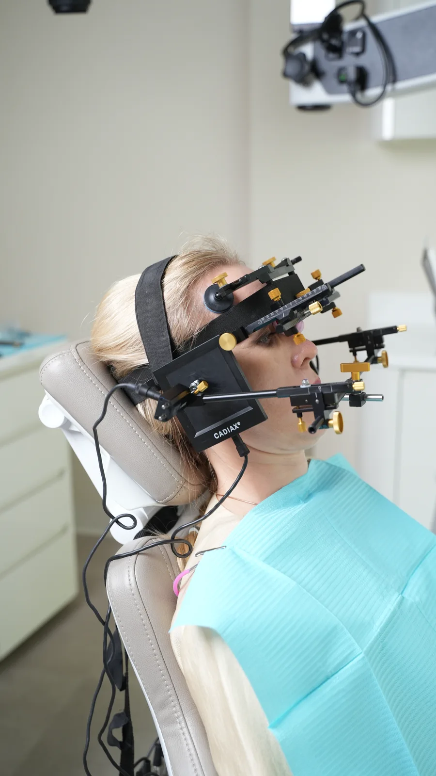
I fit the recording system and check that its attachments are secure and allow you to bring your teeth together and move your jaw freely. A loose attachment can distort the recording. I will explain any specific preparation needed before your appointment.
The assessment usually takes around 30–40 minutes, including fitting and removing the system, recording the movements and checking the results.
What happens during the recording?
I explain and demonstrate each movement: opening and closing, moving your lower jaw forward and moving it to either side. Other movements may be recorded depending on what we need to examine. Follow the instructions and tell me if a movement hurts.
Repeating movements helps assess consistency. If traces differ, we consider pain, how the task was performed and possible recording interference. I interpret repeated recordings alongside examination findings and symptoms.
Reading the traces and measurements
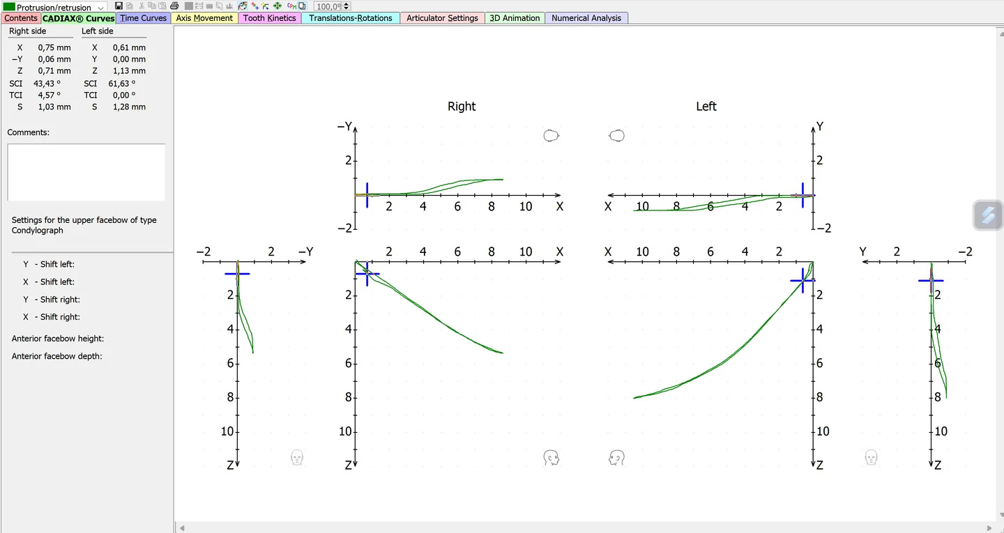
The recording shown here captures protrusion of the lower jaw and its return. Right and left sides are displayed separately. Several projections show the same path from different directions, with scales indicating displacement.
I compare the outward and return paths, differences between the right and left sides, and how closely repeated recordings match. During the consultation, I explain what I can see and how it affects the next steps in your treatment.
What we discuss after the recording
I save the recording in your clinical notes and explain the results. Together with the examination, these help me decide whether a splint is appropriate and whether MRI or CBCT would provide useful information about the joint tissues.
A repeat recording can help assess changes after splint therapy and prepare for the next stage, such as definitive restorations. We compare movements under comparable recording conditions, alongside mouth opening, muscle findings and your symptoms.
Using the measurements in an articulator
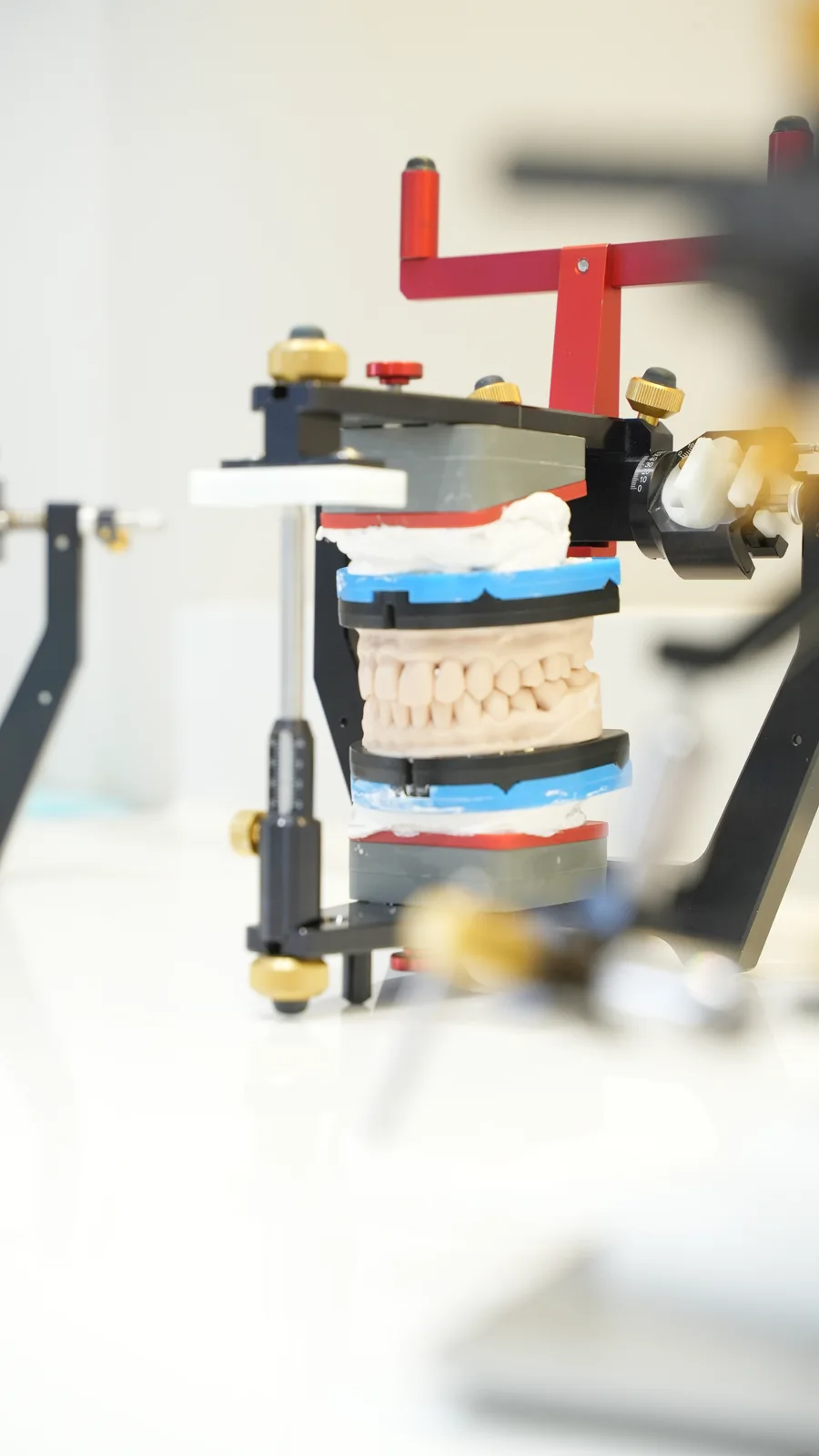
An articulator holds models of the upper and lower teeth. It helps the dentist and technician study contacts and design restorations outside the mouth. Individual movement measurements can be used to adjust its settings for the patient.
This is especially useful when restoring several teeth: we need to check how they will meet as you close and move your jaw. I then check the finished restorations in your mouth and ask how they feel. The articulator reproduces the movements set on it, but not muscle activity or the responses of living tissues.
Record
Select movement parameters relevant to the planning task.
Settings
Transfer appropriate values to articulator settings.
Design
Assess proposed surface contacts on models and then clinically.
How is this different from CBCT or MRI?
Condylography records jaw movement. Cone beam computed tomography (CBCT) shows bone, while magnetic resonance imaging (MRI) shows the disc and other soft tissues. I choose the investigation according to what needs a closer look.
In a complex case these findings may complement one another. Movement recordings, examination findings and imaging help relate joint function to your symptoms. If MRI is not possible, I plan care using the available findings and identify what needs monitoring.
| Method | What is assessed | Information obtained |
|---|---|---|
| Condylography | Lower jaw movement | Movement traces and parameters. |
| CBCT | Bone | Images of bony form and condition. |
| MRI | Disc and other soft tissues | Images of structures and their relationships in the positions examined. |